Onion Skin Cell Diagram
Cells cheek ncert microscope blotting cbsetuts cbse Onion structure biology mic microscopy Onion cell epidermal diagram labeled cells microscope under drawing skin epidermis lab bulb mag membrane observation vacuole nucleus leaves preparation
Onion Peel Cell Diagram Drawing - Djordjezivaljevic
Onion peel px Plant & animal cells staining lab answers Onion peel cell diagram drawing
Onion peel cell diagram with label
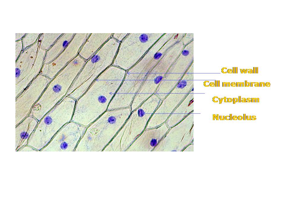
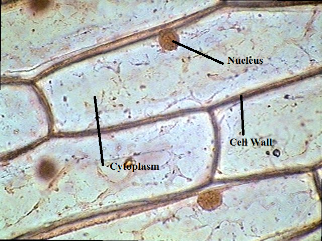
Figure of onion peel showing cellOnion cell cells skin epidermal nucleus structure microscope 100x iodine under stained shows alamy stock shopping cart high Ncert class 9 science lab manualThe science scoop: onion cell lab.
Onion skin cells (epidermal cells) shows cell structure and nucleusOnion cells under microscope Biopedia: practicalsOnion cell hi-res stock photography and images.

Onion cell peel draw cytoplasm membrane vacuole showing brainly figure
Onion microscope structure staining microscopic schoolworkhelper biology shapes15 best molecular biology visual references images Onion cells hi-res stock photography and imagesOnion cell epidermal peel size.
Magnified 40x times 100x microscopyOnion epidermal cell diagram Onion microscope epidermis 4x onionsOnion cell micrograph microscope cells stock microscopic section root allium cepa scale epidermis alamy bulb tip organelles.

Onion epidermal cell labeled diagram
Onion cell cells epidermal skin nucleus structure shows epidermis 100x stained stock alamy iodine photography live high alliumDraw the figure of an onion peel showing cell Cell onion peel vacuole cytoplasm showing figure nucleusOnion epidermal drawings epidermis labeled biology chromosomes chromosome dna observation.
Onion cell 400x lab microscope under labeled cells structure scoop science looked .

Biopedia: Practicals

NCERT Class 9 Science Lab Manual - Slide of Onion Peel and Cheek Cells

Plant & Animal Cells Staining Lab Answers - SchoolWorkHelper

Onion Peel Cell Diagram Drawing - Djordjezivaljevic

Onion Epidermal Cell Diagram
The Science Scoop: Onion Cell Lab

Figure of onion peel showing cell - Brainly.in

Onion Peel Cell Diagram With Label - itsessiii

Onion Epidermal Cell Labeled Diagram - Wiring Diagram Pictures